
Viruses for Good
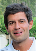
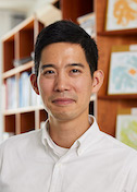
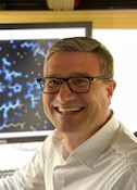
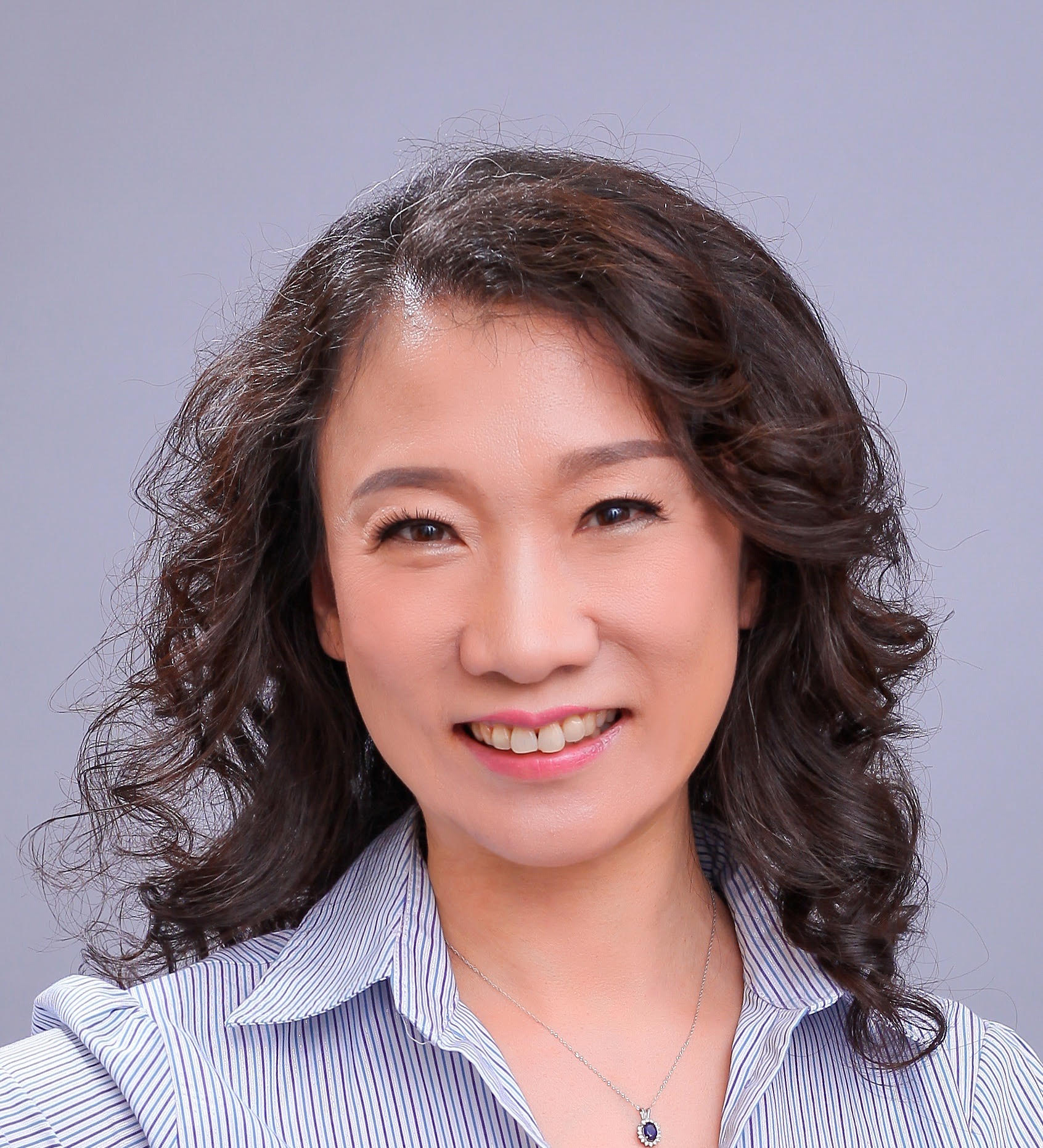
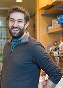
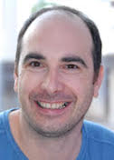
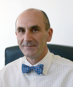
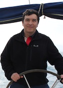
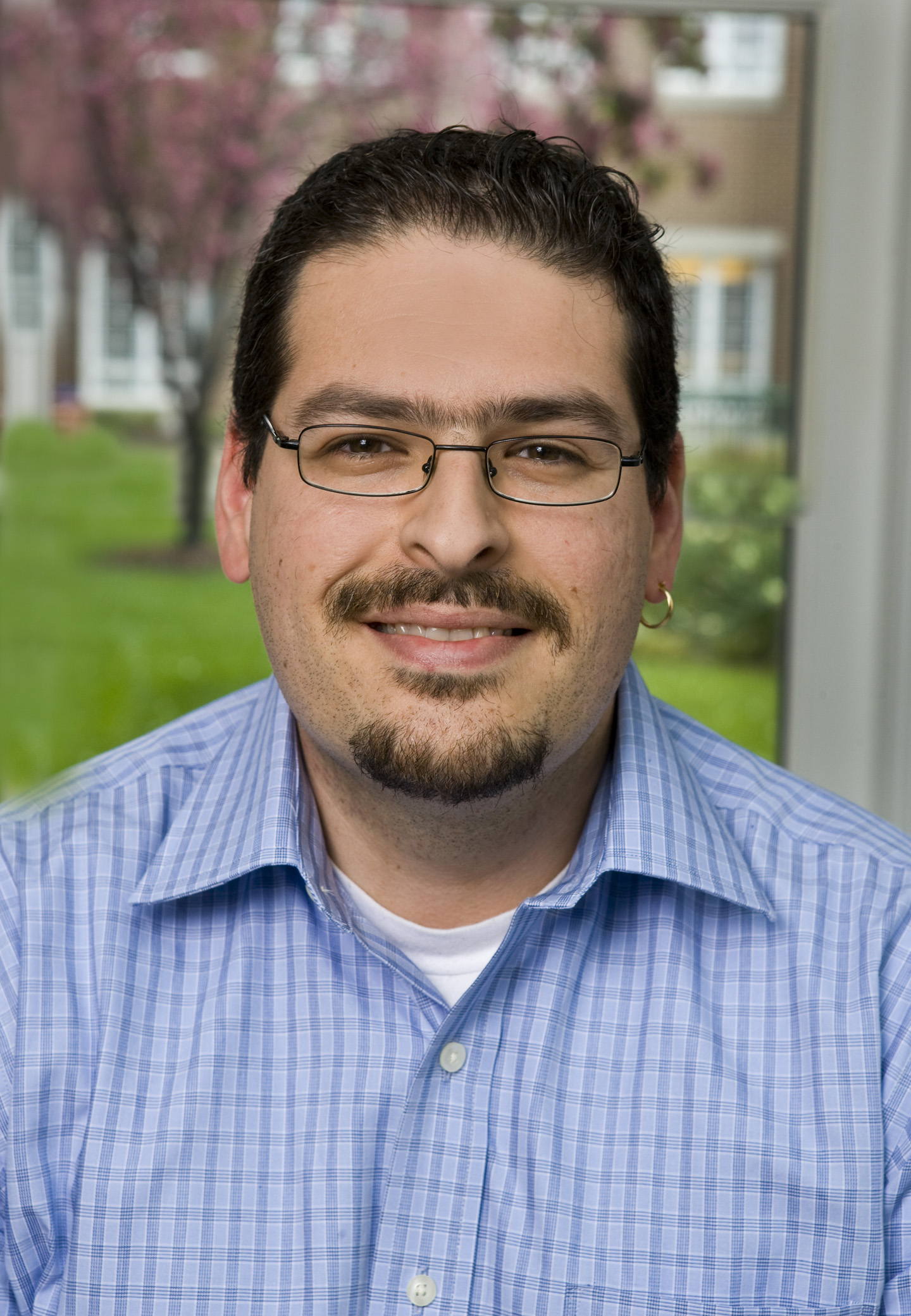
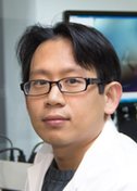
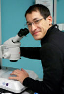
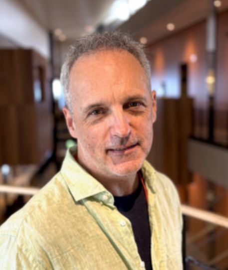
Mihnea Bostina
University of Otago
Published August 26, 2026
Viruses are usually studied as villains. After all, they can spread disease and death, disrupt society, and destroy crops. Fewer people look to them as allies. But viruses aren't all bad. Some may have the power to clear infections and fight tumors.
In his New Zealand laboratory, Mihnea Bostina wants to understand and harness the therapeutic potential lurking in some viruses to treat people and plants. "We work with a lot of viruses, mostly good viruses," he says.
One virus the lab studies selectively infects and kills certain tumor cells, while sparing healthy tissue. The group also works with bacteriophages, a type of virus that infects only bacteria, raising hopes that one day it may help overcome the problem of antibiotic resistance. For these studies, the lab deploys cryo-electron microscopy (cryoEM), which comprises about half of the lab's work.
CryoEM took center stage in structural biology methods when it broke through to atomic resolution, but the other half of the Bostina lab is zooming out with an emerging technique known as volume electron microscopy (vEM). They are exploring more life-like 3D tissue models of cancer and viral infections to gather structural information at a resolution that bridges traditional cell and structural biology. "It's better than light microscopy, but not as good as cryoEM," he says.
Virus versus tumors
Bostina has been captivated by viruses since his earliest days as a scientist. "Structurally, they have these beautiful symmetries," he says. "And it was an interesting question: How do those proteins know to self-assemble with such precision in certain conditions, and in such stable protein cages, which are almost indestructible by pH and temperature.
"They can resist a lot of chemical and physical factors, and suddenly, when the right moment occurs, the right receptor, the same solid, beautiful symmetrical structure disassembles and delivers the genome, and rapidly takes over the cells," he says.
Bostina helped identify the tumor receptor for the anti-cancer virus, Seneca Valley Virus (SVV-001), which seems most attracted to neuroendocrine tumors, such as small-cell lung cancer. The discovery of the receptor suggested a way to screen for patients more likely to benefit. A clinical study by US researchers is underway in Miami.
The lab's subsequent structural studies of SVV-001 have helped explain how the virus homes in on a receptor protein overexpressed in tumors (TEM8, also known as anthrax toxin receptor 1) and not on a similar protein in healthy cells. (PNAS, 2018) More recent studies have revealed how the virus enters the tumor cell and delivers its genome. (Journal of Virology, 2025)
In one barrier for effective treatment, oncolytic virus strategies are short-lived. "The problem with all these viruses is that the body develops antibodies," he says, "so there is a lot of interest in trying to give these good viruses a better chance to kill the tumor. We're working a bit with that."
Viruses versus bacteria
As multi-drug-resistant bacteria spread, due to overuse and misuse of antibiotics, interest in bacteriophages has grown as a possible solution. Until recently, phages were difficult to study, because they have large, complicated architectures that are difficult to decipher in atomic detail. But now "microscopes are better, software is faster, and people are more enthusiastic," he says. "Now it's a kind of avalanche of new structures."
The family of phages Bostina's team studies are the largest collection of viruses on the planet. The phages pack their genes into soccer-ball-like capsids with tail machinery that injects their DNA into specific bacteria. The capsids are made of many copies of identical proteins. The major capsid protein itself varies from phage to phage, but all tailed phages share a famous 3D fold named HK97. The identical capsid proteins adopt subtle variations on the core HK97 fold to construct and close the icosahedral cages.
The team has collaborated with researchers from overseas laboratories on several structures. They are looking at how the proteins regulate the tail injection mechanism and what prompts the sudden structural changes. They aim to build models to tease out universal principals about how phages interact with their bacterial hosts and also to understand the idiosyncrasies of specific phage infections.
One study in collaboration with Okinawa Institute of Science and Technology in Japan looked at a bacteriophage (phiTE) that infects a potato plant pathogen. The bacterium Pectobacterium atrosepticum causes stems to blacken, tissues to decay, and often leads to plant death, resulting in significant agricultural losses each year. The study provided a structural atlas of the phage and may inform rational design of biocontrol agents against plant pathogens that cause diseases such as soft rot and blackleg disease in potatoes, the team notes. (Nature Communications, 2025)
Another study looked at Bas63, a phage that infects E. coli bacteria. It is related to a phage that infects salmonella that has been engineered into a biodetection tool for detecting salmonella in food samples. Bostina and his colleagues chose Bas63 for its unique genome and were able to match protein shapes to previously unannotated genetic codes. Overall, the phage features structural elements found in three distant families of phages. (Science Advances, 2025)
"It was very interesting to find that the general architecture of the bacteriophage are in many ways identical in a lot of related viruses, but they are very, very different in the regions which recognize the receptor, which gives us hope that we can take the bacteriophage, understand the structure very well, and after that modify only a little part to make it specific to a certain bacteria."
The bacteria will mutate to resist the phage, but scientists can identify the domain and engineer a mutation that compensates and enables the phage to continue to fight the bacteria. This is something structural biologists do for all types of treatments, Bostina notes.
"We have solved so many phage viruses that we have started to understand that there are certain little domains, certain little loops, and certain little termini, which are regulating that conformational change, and there are other little proteins that are stabilizing those structures," he says. "It's an avalanche of conformational change, which has to come exactly in the right succession for the infections. They are very dynamic."
Around the World with CryoEM
Bostina grew up in Romania, the son of humanities professors. His mother specialized in Greek and Latin, and his father in French. The former communist country emphasized science over other subjects during his school years. "Basically everybody did math or physics," says Bostina, who graduated University of Bucharest with a degree in each.
He started doctoral studies in environmental science in Bucharest but reconsidered and headed to the Max Planck Institute of Biophysics in Frankfurt. He earned his PhD using cryo-electron microscopy to study membrane proteins in the lab of Werner Kühlbrandt, who later coined the term "resolution revolution." Bostina stayed for a short postdoctoral fellowship.
In 2005, he began the first of westward moves, stopping in Boston for five years to apply cryoEM to polio virus studies in the lab of James Hogle at Harvard Medical School. In 2010, he moved to McGill University in Montreal, where they installed one of the first of the new generation of powerful electron microscopes. "This was before the introduction of direct detectors, still in the 'blobology' era of electron microscopy," Bostina says.
In 2013, he moved to New Zealand to join the faculty at the country's oldest university. There he also leads the microscopy center with 21st century microscopy instruments while experiencing an academic life with the free-ranging intellectual breadth reminiscent of an earlier era.
University of Otago is located in the coastal city of Dunedin on the larger South Island, where about one-quarter of the NZ population are spread over more than half the country's land mass. The eastern tip of the peninsula that forms the town's harbor shelters the only mainland breeding colony of Royal Albatross, the world's largest birds with a three-meter wingspan. "They are by far my favorite birds," he says. The land rises quickly to the mountains where NZ mountaineer Sir Edmund Hillary trained for his historic summit of Mt. Everest.
From campus, Bostina can drive home a few miles one direction where he shares his rural property with sheep or a few miles the other direction to see a concert or sip excellent coffee at one of the cafes in town.
The small population, remote location and distinctive ecosystems combine to give research in New Zealand "a bit of the charm and the flavor of the 19th century sciences, because you always are in contact with all scientists, who are doing completely different things," Bostina says.
It's an interesting contrast to the scientific centers of Harvard, McGill and Max Planck where he trained and first worked, he says. "Of course, this is a challenge on a scientific level, because you don't interact with people who understand and can advise you on your work, but you always gain something when you lose something."
Mind the Gap
In New Zealand, Bostina became interested in volume microscopy (vEM), which "fills the gap" between light microscopy and structural biology. Dubbed the "quiet revolution" (Nature, 2023), vEM can probe the 3D structures of tissue, from organelles within cells to communities of cells within tissue. It was originally used to examine connections and activity of neurons in the brain.
"We're trying to understand the ultrastructure of biopsies of cancer," he says. The team is working with 3D cultures of cell lines and with patient biopsies. Ultimately, they want to know why some of the same cancers in different people respond to the same treatment, while others do not. He predicts the vEM data will supplement other clinical diagnostic and prognostic measures, such as genomics and proteomics, and also be useful in drug discovery.
In a proof-of-principle study, his team conducted a comprehensive analysis of the ultrastructural morphological differences between cancer cells cultured as 3D tumorspheres and those cultured as 2D monolayers. The samples were frozen, fixed and then prepared to be imaged with a scanning electron microscope. The tumorsphere model more accurately represented the in vivo tumor structure, they found, including a diverse array of mitochondrial morphologies exclusively in the 3D culture. (Journal of Structural Biology, 2023)
Bostina compares vEM's development stage to cryoEM before the resolution revolution. Frozen samples are sliced and imaged by a scanning or transmission electron microscope. They don't have to be labeled. Then software must be trained on features of interest, such as mitochondria or chromosomes. vEM now involves a piecemeal workflow, with multiple formats that require user skills to get the data from one part to another.
The Bostina lab has made its software available on SBGrid (Volume Segmentation Tool (VST). He cautions that software trained on one sample doesn't automatically work on another. "Brain cells are very different from stomach cells and very different from skin cells," he says, adding that AI methods are not very powerful yet for vEM.
"There's a lot of space for people to develop tools," he says, a lot of interest in making the software faster, better and more specific to certain cells."
------------By Carol Cruzan Morton
Carol Cruzan Morton is a senior science writer and medical editor in Oregon. She contributes profiles to SBGrid and has also written for Science,
Medscape, Oregonian, Boston Globe, and San Francisco Chronicle, among other publications.