The Shapes of Energy
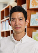
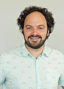
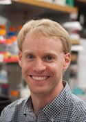
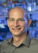
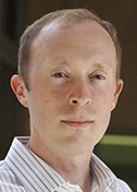
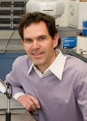
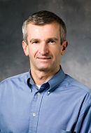
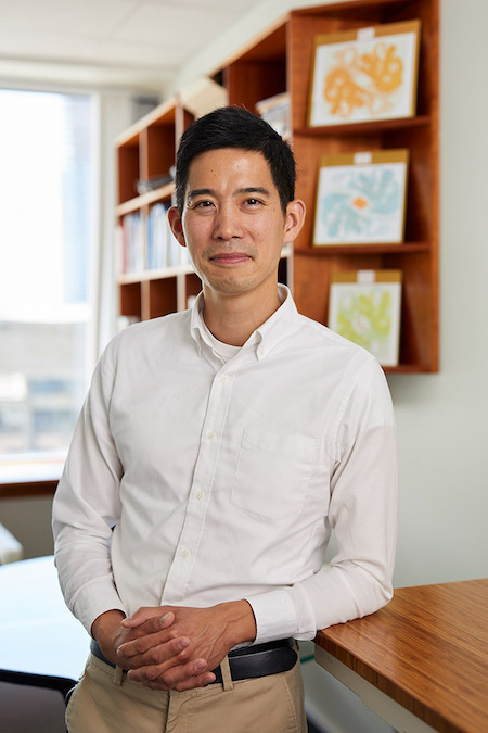
Luke Chao
Massachusetts General Hospital
Published December 12, 2024
In Woods Hole, Mass., the Atlantic breeze mixes with the hum of microscopes and the chatter of biologists. There, in a summer neurobiology course, Luke Chao first became enthralled by mitochondria “whizzing” around the neurons in a mouse.
“It was just really cool,” he recalls, describing the tiny organelles as they traveled along the long projections of nerve cells (axons), in a model of neurological damage.
This inspirational moment has since grown into the focus of the Chao Lab at Massachusetts General Hospital (MGH) in Boston. There, the Chao team explores the dynamic and enigmatic architecture of cellular membranes, particularly those of mitochondria, which provide energy for the cells in our bodies.
Two membrane layers form mitochondria, but the hub of energy production is the wrinkled inner layer folds, called cristae, which are distinctive for their intricate and diverse shapes.
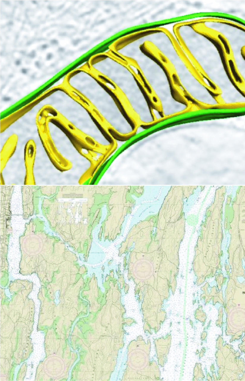
“The folds can come in tubular forms,” Chao says. “Or they can come in sheets. One can describe them like pasta geometries — lasagna (lamellar, or sheet-like), or penne (tubular). It has remained a mystery how the different forms are made, and how the different shapes facilitate the organelle’s varied functions.”
Chao has had an interest in the classic structural questions about the atomic arrangement of individual proteins that sculpt mitochondria. But he’s also interested in the bigger picture — how multiple proteins come together in unique and special combinations to build membrane architectures in cells.
“We'd like to understand on a very fundamental level how cristae are made,” he says, “to understand structure-function relationships of how proteins influence membranes.” Understanding how shapes change under different conditions could shed light on diseases such as neurodegeneration, cancer and aging, where mitochondrial dysfunction is an early and important player.
Chao grew up in Chelmsford, Mass. During his undergraduate years at Brandeis University, he took a work-study job washing dishes in a biochemistry lab. “I just loved the environment,” he says. “Everyone working together, fumbling in the dark trying to figure things out.”
Chao went on to pursue a Ph.D. at the University of California, Berkeley, under the mentorship of structural biologist John Kuriyan. There, Chao developed a fascination with the “amazing acrobatics” of proteins, uncovering their roles as dynamic machines that reconfigure themselves to perform vital cellular functions.
His doctoral work focused on the calcium/calmodulin-dependent protein kinase II (CaMKII) holoenzyme, a large oligomeric molecule central to neuron and cardiac calcium signaling. His findings revealed mechanisms of protein activation and inhibition, illuminating how these molecular giants operate with precision in living cells.
Postdoctoral work in the laboratory of Stephen Harrison at Harvard Medical School introduced Chao to the realm of membranes and viruses, specifically flaviviruses, a family that includes Dengue, West Nile and Zika. There, he investigated the conformational changes that proteins undergo to mediate membrane fusion—a process crucial in viral entry.
Toward the end of his postdoc, Chao pondered his next move. He recalled his experience with mitochondria at the Marine Biology Laboratory in Woods Hole and saw new opportunities to explore protein acrobatics and membrane biology.
“To me, it’s a fascinating medium for investigating shape and motion across scales,” he says. “Obviously, there are the individual lipids, their position, their composition—all of these details matter to generate the right environment for the proteins to work. But the proteins themselves also act on the fluid bilayer to generate highly specialized local environments. There are fantastic opportunities for integrative understanding of these relationships at multiple levels. We still do not understand how all of these factors work together to generate a living, breathing organelle.”
In 2016, Chao launched his independent lab at MGH, diving into the dynamic architecture of mitochondria. On one level, the Chao group studies mitochondrial fusion with in vitro techniques, similar to the viral fusion experiments of his postdoctoral training, reconstituting essential components to answer detailed mechanistic questions (Ge et al., eLife 2020). By isolating proteins and recreating the membrane environments in the lab, Chao can dissect membrane fusion and protein-membrane intermediates.
On another level, his work leverages techniques such as cryo-electron tomography (cryo-ET), which allows researchers to freeze cells and capture 3D snapshots of their inner workings. It’s a way to see structures “in their native context,” he notes, avoiding the artifacts introduced by methods that rely on fixation and staining.
An area of focus for Chao’s lab is the protein Opa1, which reshapes mitochondrial cristae in response to stress. In a recent study, Chao and his team examined how Opa1’s state alters cristae structures. The findings revealed that mitochondrial membranes can adapt dynamically, reshaping themselves to meet the energy demands of the cell or to respond to environmental challenges (Fry, Navarro, et. al, EMBO J, 2024).
Chao also looks at these questions through an evolutionary lens. The origin of mitochondria has been dated to over a billion years ago—a time when primordial archaea absorbed an alphaproteobacteria, the mitochondrial precursor.
“There are bacterial analogues of cristae, called intra-cytoplasic membranes,” he says. “The common theme is that they help provide additional real estate to pack in different biosynthetic enzymes, similar to how the expanded surface area of the mitochondrial inner-membrane packs in a lot of respiratory chain proteins. So there might be some conserved principles in how they are built, and some interesting differences too.”
In collaborations, he partners with MGH colleagues, labs in the Boston area, as well as groups internationally. Locally, his home Department of Molecular Biology at MGH has “people doing all sorts of interesting things, from prokaryotic immunity and cilia dynamics, to RNAs, plant and chromatin biology,” feeding his intellectual curiosity.
Outside the lab, Chao and his family often escape to explore the mountains of New England and enjoy Maine’s rugged coastline, a place reminiscent of mitochondria cristae. “There are so many inlets that if you take the tidal coastline of Maine, its actually greater than that of California. It’s all about surface area. There is a lot to explore.”
--Carol Cruzan Morton

