Reshaping Membranes
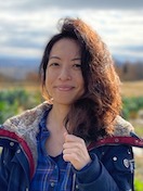
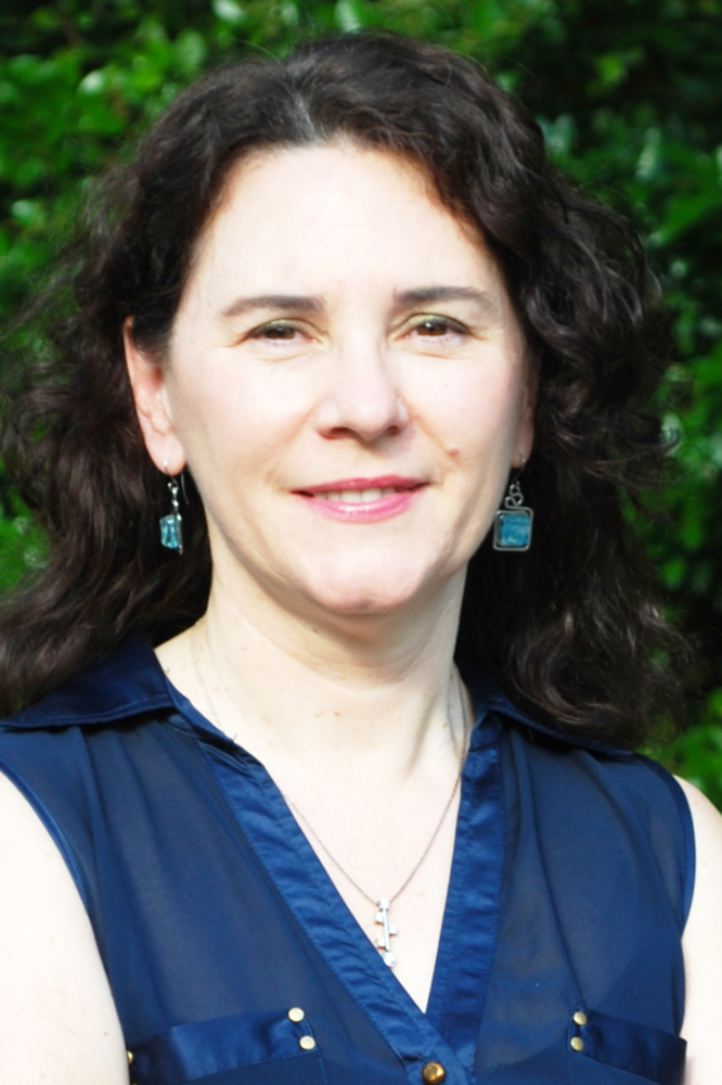
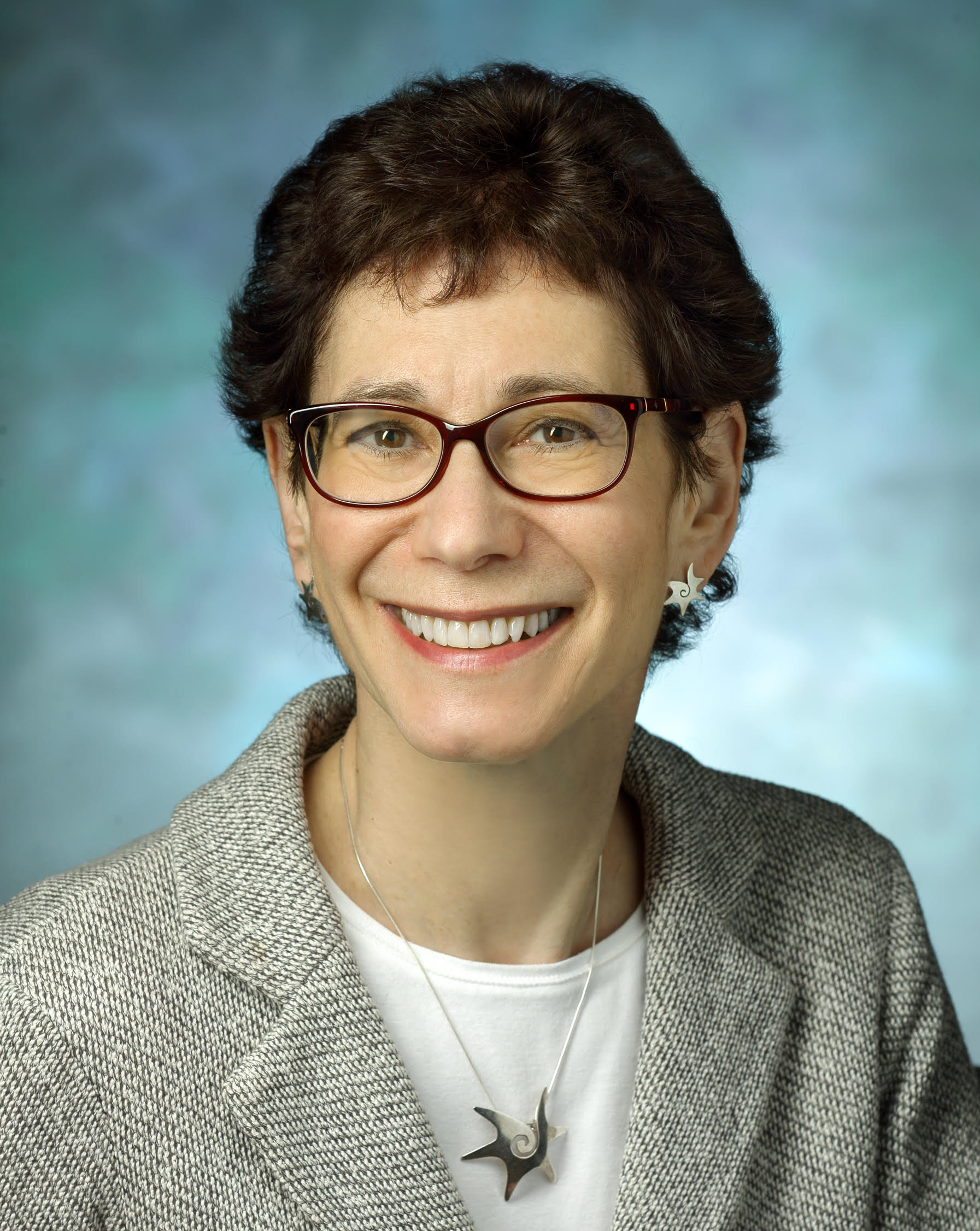
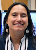
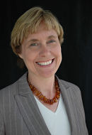
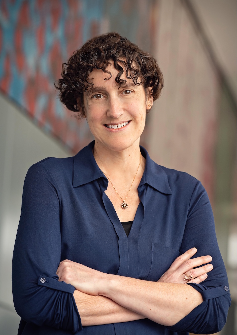
Melanie Ohi
University of Michigan
Published November 23, 2025
Cryo-electron microscopy has been a boon for the study of membrane proteins. No crystallization needed, and the ability to visualize dynamic regions of the protein to boot. But one aspect Melanie Ohi especially likes about single particle EM is the immediate gratification of seeing even the blurriest pictures.
“You don't have to wait for the punch line, because you can actually see an image,” Ohi says. “You don't know the structure right away, but you can see some things quickly, such as: What is the overall shape? Are there parts that are flexible? Does it just look horrible, so that you immediately know the purification strategy has to be improved?”
The Ohi lab at the Life Sciences Institute at the University of Michigan (U-M) determines detailed three-dimensional structures of membrane proteins that work together as complex molecular machines and that are important in health and disease.
These days, cryo-EM can capture the atomic architecture of complexes, but in her lab the sharper details often come at the end of a long research process that requires the investigation of other relevant biology along the way.
“We can answer questions that take advantage of the entire gamut of the resolution range of electron microscopy,” Ohi says. “I like using the whole range. We can learn something at 15 angstroms, and we can learn something at 3 angstroms. It's just a matter of what kind of questions you want to address.”
Those questions range from the secretion systems pathogenic bacteria use to breach membrane barriers of cells to cause disease to how blood clotting is initiated and regulated.
The broad interests of the lab are connected by an overarching theme. “I like to understand how large protein complexes function—how different pieces come together to synergize and to do something that one protein alone is not able to do,” Ohi says.
On a macro level, Ohi follows the same principle by working in collaborations with other labs. “There're lots of ideas, but actually getting them to work is very hard,” she says. “It’s more fun to work with a group of people from different disciplines who are willing to get into the details of the hard process of how best to purify samples.”
Moving Across Membranes
To infect people, bacteria have devised ways to move molecules, such as virulence factors and DNA, into the host cell. Ohi and her colleagues were the first to purify and determine high-resolution structures of the most versatile and complicated of these complexes, called type IV secretion systems (T4SSs). They looked at the T4SSs of two kinds of bacteria that use different strategies to cause disease.
T4SS enables Legionella pneumophila, for example, to unleash more than 300 different proteins into the host cell. The bacteria cause Legionnaires’ disease, a lung infection that targets macrophages, a type of white blood cell, and often spreads to people by contaminated water systems.
In contrast, once Helicobacter pylori have colonized the stomach, they inject a single protein, CagA, into the endothelial cell lining. The protein, the only known bacterial oncogene, is present in some strains and not others. It triggers chronic gastric mucosa inflammation and is a strong risk factor for ulcers and stomach cancer.
“We were hoping that if we determine the structure of both T4SSs, we would see structurally how one complex translocates one protein across the membrane, while the other complex can translocate hundreds of proteins,” she says, but “no, not yet. There's still more work to be done, but there are some key similarities and differences that we are now exploring.” The project is a long-term collaboration with H.pylori expert Tim Cover MD and members of his lab at Vanderbilt University Medical School.
The Structural Journey
Ohi grew up in Spokane, a city in Washington near the Idaho border. Her mom was a teacher and her dad a civil engineer. She moved 300 miles west for college to Pacific Lutheran University near Tacoma, thinking her aptitude for math and quantification would be a good fit with physical chemistry.
Her first biochemistry class changed her trajectory. Maybe it was pictures of molecular structures she saw in class, but for reasons she can’t remember, she liked the idea of becoming a crystallographer and soon decided to go to graduate school.
“I didn’t grow up in a scientific family,” she says. “My family was like, what are you doing? Because it’s not a job, right? This is where I really got lucky.”
Perusing scientific journals in the library, she read about a crystal structure of a cyclin-dependent kinase, a family of molecular switches that drive cell division and growth. One name popped up repeatedly, Kathleen Gould at Vanderbilt University. When Ohi interviewed on campus, she was surprised to learn that Gould was not a crystallographer but a yeast geneticist.
In her first year, she rotated through different labs. She sampled a crystallography lab and didn’t like the technique as much as she thought she would, “because you work very hard to get crystals so you can determine a structure, but if you don't get crystals, you can't answer a question,” she says. “And that part really bothered me.”
Accidental Geneticist
But when Ohi rotated into Gould’s lab, it felt like a good fit. And it set the stage for how she runs her own lab.
“Even though it was an uncomfortable intellectual experience, because I didn’t know genetics or anything about yeast, I really liked the way that Kathy thought about science and the way that she posed questions,” Ohi says. “I thought it would be good to learn how to do hypothesis-driven research. In crystallography, it seemed like you actually had to solve the structure before you could make a hypothesis, which is fine, and that's the way it works. But I didn't want four years of getting the crystal and then leaving before I could test the hypothesis.”
Luckily Gould was willing to take a chance accepting a student with no biological experience. In her training as a yeast geneticist, Ohi learned how to tag and purify proteins from the endogenous source, an approach that paid off later for isolating T4SSs in her own lab years later. “We allow the organism itself to put the complex together the way it wants to, even though it can be very challenging to purify enough of it for structural analysis,” she says. In the case of Legionella, this approach allowed them to find components not previously identified as part of the T4SSs.
She also absorbed a new perspective. “People who are not structural biologists only get excited by atomic resolution structures when they provide insight into function,” she says. “They get really bored when you start to talk about atomic details of a structure unless those details provide biological insights. However, if you can show where regions of a protein or complex move—like this whole domain swings to a different position when activated by another enzyme, or a structure changes conformation when proteins interact—that gets exciting for everyone.”
By the time she was finishing up graduate school, cryo-EM was emerging as a viable technique for determining 3D structures of large proteins and complexes. She noticed papers reporting structures of multiprotein complexes that couldn’t be easily crystallized or were too big for nuclear magnetic resonance (NMR) analysis.
Beginning Blobology
“I still wanted to be a structural biologist,” says Ohi, who defied prevailing advice to choose other structural approaches over cryo-EM. “Most of my mentors warned me that if you do a post-doctoral fellowship using cryo-EM, you will likely need to do a second postdoctoral fellowship, because EM is a niche technique that will never routinely achieve high resolution structures. You will be a “blobologist,” and it will be hard to find a faculty position, because the equipment is so expensive, and you'll struggle to get to resolutions that answer mechanistic questions. And their advice was absolutely correct at that point in time.”
But she had just spent five years in a cell biology department, and she sensed that many scientists would value even 2D EM images of dynamic complexes. If the questions are interesting, images—even lower-resolution ones—can be powerful. And, not for the first time, she reasoned that it wasn’t the end of the world if it didn’t work out. In the here and now, it was what she found interesting.
She took a leap and landed a postdoctoral fellowship in the lab of Thomas Walz, then at Harvard Medical School (now at The Rockefeller University). As it turned out, “that was one of the best decisions I ever made,” she says. “Tom was a great scientific mentor.” She learned single particle EM from Walz and others in his lab while tackling structurally challenging projects. She embraced the idea that compelling biology can be uncovered even without atomic-level details—a philosophy that still guides her lab.
Putting it all together
Ohi joined the faculty at Vanderbilt in 2007. She ran a lab there for a decade, as advances in cryo-EM technology and techniques enabled higher resolutions sufficient to build models with atomic details. In 2017, she joined the faculty at University of Michigan in Ann Arbor, where she set up a lab in the Life Sciences Institute and as a member of the Department of Cell and Developmental Biology in the medical school. At U-M, her lab continues to work in long-term multidisciplinary collaborations and on projects that require creativity in preparing samples for structural analysis.
One of these collaborations is with Anne Kenworthy at University of Virginia and involves Caveolin-1, a protein building block of caveolae, which are small cave-like invaginations localized at the plasma membrane. Caveolae are found in various cell types, such as muscles, epithelial cells and fat cells, apparently in service of a wide range of tasks yet to be completely understood by scientists, although their abnormal functions are implicated in cancer and cardiovascular disease. Kenworthy also recently discovered that caveolins are found across a broad evolutionary spectrum, including organisms that likely do not form caveolae at all, leading to more questions about the functional roles of caveolin proteins.
Caveolin-1 is a membrane protein that dips in and out of the lipid bilayer but doesn’t cross it like a conventional transmembrane protein. “It was hard to find the right way to purify and then vitrify Caveolin-1 for cryo-EM analysis,” says Ohi.
Once Ohi and Kenworthy saw the 3D structure of Caveolin-1, they immediately knew it would challenge dogma for how this protein interacts with and shapes membranes.
“So now we are trying to figure out exactly how does it bend the membrane,” she says. “Our structure suggests a new model for how caveolin inserts into membranes, Now that we have a structural blueprint, we can design very specific mutations for doing structure-function analyses combined with cellular experiments that explore what is structurally important for caveolin’s roles in signaling and what regions of the protein are important for its membrane bending.”
In another project, the Ohi lab teamed up with U-M colleague Jim Morrissey, an expert in blood clotting enzymes, to look at protein complexes that assemble on membranes to initiate the blood clotting enzymatic cascade. “The tissue factor is on the surface of cells and the proteins that are involved in initiating the blood clotting are in our blood, and there's always a question of what keeps them from either initiating or not initiating blood clotting, since mistakes made in either direction of regulation cause major health issues,” she says.
One of the triggers that initiates the blood clotting cascade is a charged lipid that flips from the inside to the outside of the cell in response to cellular injury and ultimately leads to the activation of a protease called Factor X. So Ohi and her colleagues built a protein complex that includes three key proteins on a nano disc composed of charged lipids and determined the cryo-EM structure. They found that a very small movement—four or five angstroms—in one of the proteins puts a Factor X domain in the right position to be activated. (Blood, 2025)
Lab philosophy
To unwind, Ohi enjoys gravel cycling and belongs to a cycling club. To avoid run-ins with student drivers, she prefers to walk the mile or so between work and home, but she rides on less trafficked gravel roads around Ann Arbor whenever she gets the chance. “I don’t think I’m a very competitive cyclist, but I’m definitely a very serious one,” she says.
Ohi acknowledges that it’s not an easy time to be a scientist, with science feeling under attack. “I like to encourage the people in my lab to focus on the things that they can control, which is doing good science,” she says.
“I also like to tell my trainees that this job is very special. We get to work on questions that interest us, interact with very smart colleagues, and we have a lot of flexibility for how we organize our time. Since we are always learning new things, it doesn’t get stale. Even when its stressful, it’s still a pretty sweet deal to have the chance to help discover the molecular details that drive cell function.”
By Carol Cruzan Morton