Escape from the Darkroom
Wolfgang Kabsch and XDS
Max Planck Institute for Medical Research
Published May 19, 2011
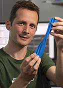
As Wolfgang Kabsch headed for the darkroom, facing another day of developing films of Xray diffraction patterns, he passed by a new machine sitting on a bench, unused. It was the mid-1980s and the machine was an early electronic Xray detector, full of new technology but lacking the software to make it usable.
“It was just sitting there, looking at me,” says Kabsch, staff scientist emeritus in biophysics at the Max Planck Institute for Medical Research. “I decided rather than wasting my time in the darkroom, I could program the detector to do something useful.”
Kabsch's efforts led to the development of XDS, Xray Detector Software. First released in 1986, the tool is still widely used today to translate raw Xray data from a variety of detectors into standardized data that includes the H, K and L indices of reflection, the intensity of the reflection, and the standard deviation of the intensity.
At the time, Kabsch was working on his main project, solving the structure of the muscle protein actin. After 14 months of exploring the detector and coding all of its features into a Fortran-based software tool, he was able to solve the actin structure with the excellent data from the new detector, rendering the film-based approach obsolete. “It was a lot of work,” says Kabsch, “but when it was done, the structures just came out like nothing.”
Kabsch released XDS in 1986 and published the actin structure in Nature in 1990. He continued to use the detector for other projects, such as solving (in collaboration with SBGrid member Emil Pai, biochemistry professor at the University of Toronto) the RAS p21 protein, an oncoprotein in human cancer.
Since that first software release, Kabsch has run XDS as a one-man shop. As new detectors come on the market, he extends the software to support them. In all, the software supports over 22 different detectors.
Recently, Kabsch added support for a novel pixel detector called PILATUS from the Paul Scherrer Institut in Switzerland. This data-intensive detector delivers ten images per second, each 6 megapixels in size. On the docket is support for a new types of detectors assembled from may separate, planar segments for recording FEL (free electron laser) data at the Linac Coherent Light Source at Stanford.
Kabsch, now retired, is always looking out for ways to keep XDS up to date. “When I see a better algorithm or a better way of computing, I put it in,” he says. He announces improvements on the XDS web page. Kabsch has also contributed to other applications, including the development of a dictionary of protein secondary structure, used in applications such as Procheck.
While Kabsch remains the primary coder of XDS, he has tapped a successor. Kay Diederichs, professor of protein crystallography and molecular bioinformatics at the University of Konstanz, has worked with Kabsch and XDS for over two decades and will continue to evolve XDS when Kabsch starts taking his retirement more seriously.
– Elizabeth Dougherty

