
Proteins at Work
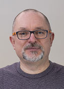
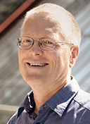
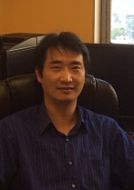
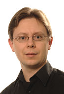
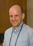
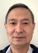
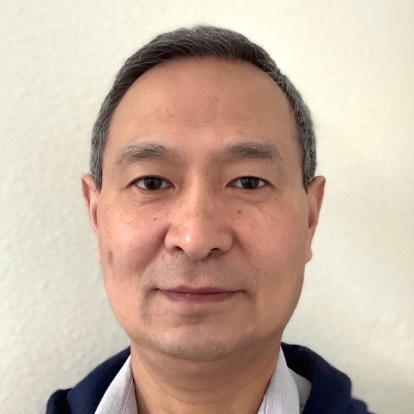
Shawn Zheng
Biohub
Published September 27, 2026
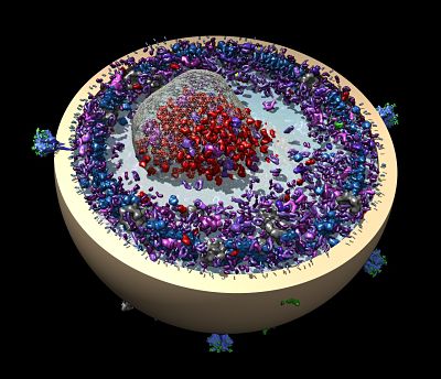
Protein databanks are bursting with 3D structures that reveal molecules in vivid atomic details. But most of these molecules have been purified and studied in isolation, outside their native cellular environments. What researchers want to see now, in comparable detail, is how these molecules live and work together inside cells.
“The context-rich structural information, with molecular detail, is a cornerstone,” says software developer Shawn Zheng. “It is essential for understanding how a cell functions at the molecular level.”
Based in Redwood City, California, Zheng leads an algorithm development team at Biohub, a nonprofit research organization launched by the Chan Zuckerburg Initiative in 2016. As its name suggests, Biohub brings together biologists, physicists, microscopists, and software and hardware engineers in a focused effort to develop breakthrough imaging technologies and methods.
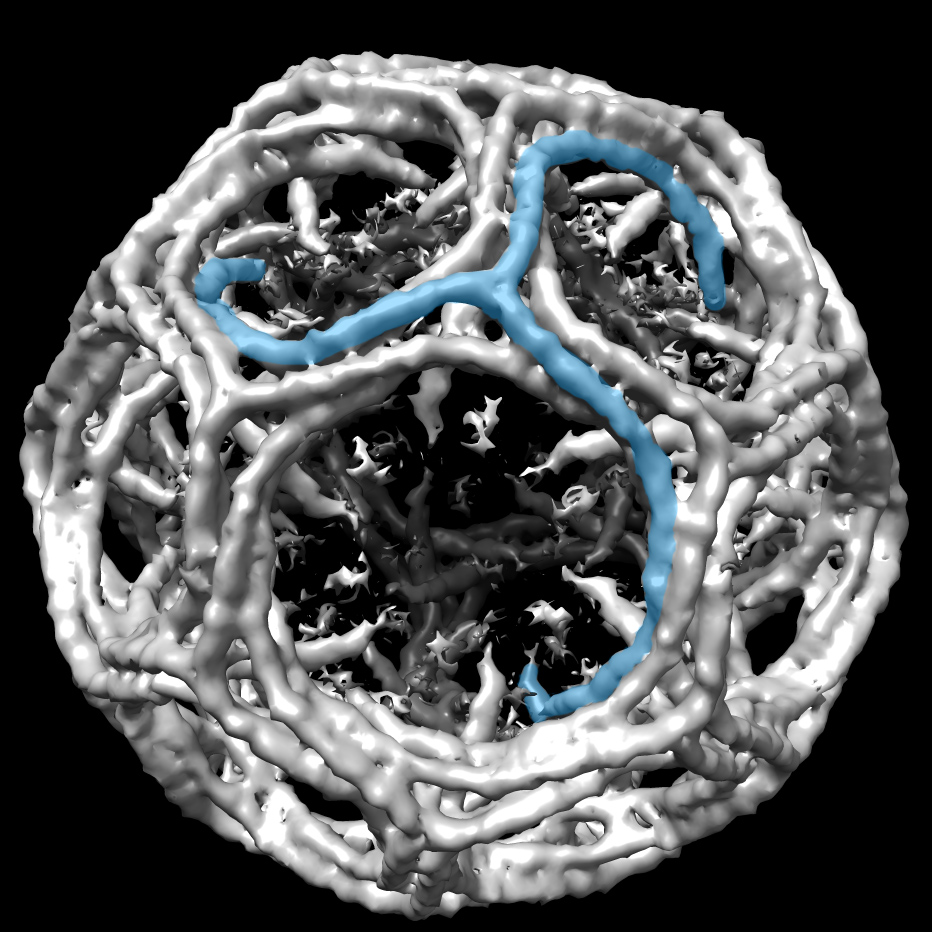
Zheng and his colleagues focus on cryo-electron tomography (cryoET). The advanced imaging technique enables scientists to visualize the three-dimensional (3D) structures of cells and biomolecules at near-atomic resolution.
In fact, they predict cryoET will likely deliver the next major advance in structural and cellular biology. The push to see molecular details in their native environments comes on the heels of the resolution revolution of single-particle cryo-electron microscopy (cryoEM).
“CryoET not only reveals the high-resolution structures of biological molecules, but also preserves their original cellular context,” he says. “It feels like we’re on the eve of another revolution.”
Challenges Abound
Their computational efforts must overcome a harsh physical reality: CryoET seeks to reconstruct high-resolution 3D structures from noisy, incompletely sampled data from thick slices of biological samples that have been flash frozen to preserve a close-to native state.
These challenges begin as the microscope bombards the biological samples with a high-energy electron beam while gradually tilting them through a range of angles. CryoET captures a tilt series of 2D images to reconstruct a 3D volume, or tomogram.
Beam-induced motion and radiation damage further complicate this task. Low electron dose settings can limit radiation damage. Even so, beam-induced motion still deforms the sample and blurs the images, while imperfections in the microscope’s optics further distort the images.
Another challenge arises from a limitation in sample tilting. At higher angles, radiation damage accumulates, further reducing the high-resolution structural information needed to interpret molecular mechanisms. As a result, cryoET tilt series are typically collected over a limited angular range, leaving a gap known as the “missing wedge” in the data used to reconstruct a tomogram.
Building Better Tomograms
In their first two years at Biohub, Zheng’s team developed a computational solution to build better tomograms (Nature Methods, 2026). Called AreTomoLive, the software package enables fully automated, comprehensively corrected, and denoised cryoET reconstructions in real-time and at high throughput.
AreTomoLive cuts the processing timeline from several months to several days, or about 80 times faster according to an in-house estimate by Biohub biologists.
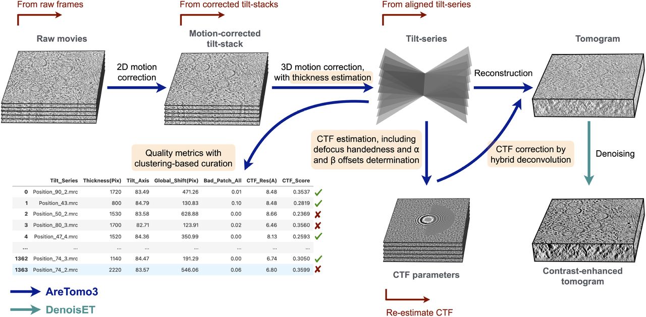
Downstream processing can begin immediately rather than waiting for acquisition to finish. The integrated pipeline also eliminates the bottlenecks of moving data between software packages and formats for different preprocessing steps.
An experimentalist can put their sample on the microscope, identify all the positions they want to collect, start AreTomoLive, and walk away without having to worry about it, says Ariana Peck, a data scientist on Zheng’s team who led the AreTomoLive development.
AreTomoLive is composed of two GPU accelerated software packages seamlessly integrated together. The first, AreTomo3, reconstructs tomograms in real time with a lot of corrections built in for all the wacky things that happen in collecting data on biological samples on an electron microscope. It extends earlier software first developed by Zheng and colleagues at University of California, San Francisco, including the MotionCor2 and AreTomo series.
The other package, DenoisET, runs in parallel with AreTomo3. It implements the canonical Noise2Noise algorithm, a statistical insight that a denoising network can be trained using only noisy image pairs.
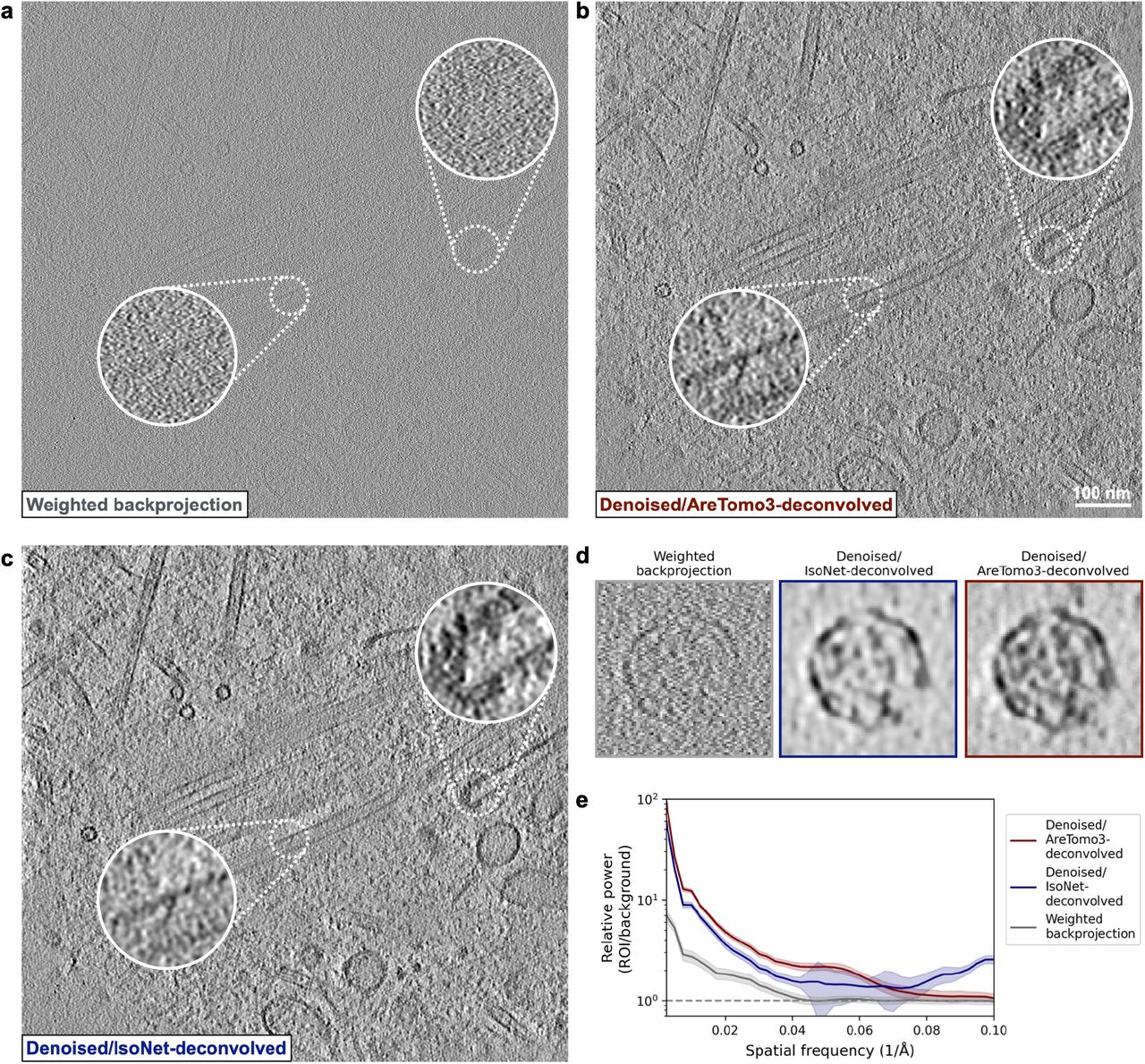
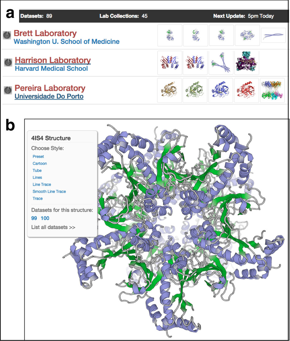
AreTomoLive can keep up with recent advances in faster parallel data collection schemes. The two packages, AreTomo3 and DenoisET, are supported by SBG, with a webinar presentation by Peck available at https://sbgrid.org/software/titles/aretomo3.
For softwear users, Zheng fields ongoing questions and requests about features they would like to see and how to get the best performance from AreTomoLive for their data set and experimental setup.
“It’s a good collaboration with the community,” he says. “Many times, the questions are inspiring. From a purely software development perspective, they may present a new use case for your software and are very insightful.”
Particle Picking
The Biohub team has moved downstream to the next data processing hurdle in the cryoET workflow: to identify tens of thousands of copies of the target molecule in a crowded cellular environment across tens to many hundreds of tomograms.
It remains prohibitively difficult to find most protein species in the 3D reconstructions, a process known as particle picking, annotating or labeling. The task is plagued by a high false positive rate and noise artifacts that can smear the original structures, Zheng says.
They are exploring a range of machine-learning strategies to identify identical molecules across different conformations while distinguishing them from look-alike molecules of other species, known as dirty picks or false positives. Zheng and his colleague jokingly coined the term “computational purification” to separate clean picks from dirty picks. They now call the approach “computational distillation.”
In the overall cryoET workflow, particle picking sets up the final stage. Once the molecular targets have been identified, the target-rich parts of the tomograms are singled out for the high-resolution step of subtomogram averaging.
Subtomogram averaging (STA) is used to recover high-resolution structural information lost to the missing wedge. In this approach, researchers identify many copies of the same molecular complex across hundreds or thousands of tomograms and computationally align and average them. By combining information from large numbers of particles in different orientations, STA can compensate information missing from individual subtomograms while greatly enhancing the signal-to-noise ratio of the resulting maps.
Casting a Wide Net
Ultimately, biologists’ needs are the driving force behind the technology development, Zheng says. “But we also want to harness the collective brilliance of the community to advance these technologies, particularly against the backdrop of the dazzling advances of AI and machine learning.”
In 2025, Biohub launched a global competition to stimulate new machine learning algorithm development for particle picking. Hosted on Kaggle, the three-month challenge galvanized a large machine learning community, attracting more than 1,000 participants from 76 countries who submitted 28,000 solutions. (Nature Methods, 2026)
The data set for the contest was released on the CryoET Data Portal (https://cryoetdataportal.czscience.com/), built in 2024 by a Biohub team to foster machine learning development using standardized data. The contest data, along with data contributed by in-house biologists, were reconstructed and denoised using AreTomoLive.
As cryoEM advanced in the 2010s, ribosome datasets became well known gold standard test cases for ever-sharper resolutions. Likewise, ribosomes are so commonly used to validate new cryoET methods at Biohub that the team jokes about “ribosome fatigue,” Zheng says, even though cryoET undoubtedly has much to reveal about this well-studied protein-building machinery in its native cellular context.
Scientific Journey
Zheng grew up in southeast China. He recalls starting school early and being the youngest kid in his class from elementary school through college. His father was a professor of material science at a local university. Zheng cites his father’s logic-based reasoning in conversations during his teenage years as a big influence that stoked his interest in a computation-related career.
After college, Zheng pursued a graduate degree in the United States. He earned his PhD in mechanical engineering from the University of Utah in 1999, focusing on the physics of fluids. He soon encountered a lean job market in experimental physics, prompting him to broaden the search to computational programming, based on the image processing skills he had developed through his physics experiments.
The first time he ever saw an electron microscope was in his job interview at UCSF with David Agard, who had an opening for a bioinformatics specialist. Agard became Zheng’s mentor for more than 20 years—longer than many marriages, Agard once joked.
At UCSF, Zheng supported Agard’s innovations in electron-counting detectors and beam-induced motion correction, advances that helped push cryoEM to atomic resolution. Despite these contributions to cryoEM, Zheng describes his career as being centered on cryoET. At UCSF, he also tackled the broader data processing challenges in cryoET, aiming to lower barriers for routine use by the nonexpert users, first with UCSFTomo and later with the MotionCor2 and AreTomo series.
Agard was the founding scientific director of what was then called the Chan Zuckerberg Imaging Institute, launched in 2022 and brought under the Biohub umbrella in 2025. He invited Zheng to join.
Zheng was drawn to the institute’s focus on developing new technology and new methods, its highly collaborative and supportive environment, and its in-house and extended network of expertise across technology fields. He takes additional inspiration from colleagues in different fields at Gordon Conferences and microscopy meetings. The Biohub further fosters this exchange through its residency program that brings together faculties and their graduate students and postdoctoral fellows from labs across the country and around the world, who regularly participate in online and in-person meetings on cryoET methods development.
Outside the lab, Zheng enjoys hiking with his wife and reading books about how high-achieving scientist leaders navigated difficult situations, such as Richard Feynman. He has a narrow range of favorite foods, yet many of which are easily available in the Bay Area, including fish and chips.
-----------------------------By Carol Cruzan Morton
Carol Cruzan Morton is a senior science writer and medical editor in Oregon. She contributes profiles to SBGrid and has also written for Science, Medscape, Oregonian, Boston Globe, and San Francisco Chronicle, among other publications.