The Final Phase
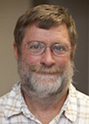
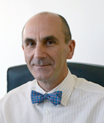
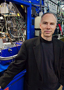
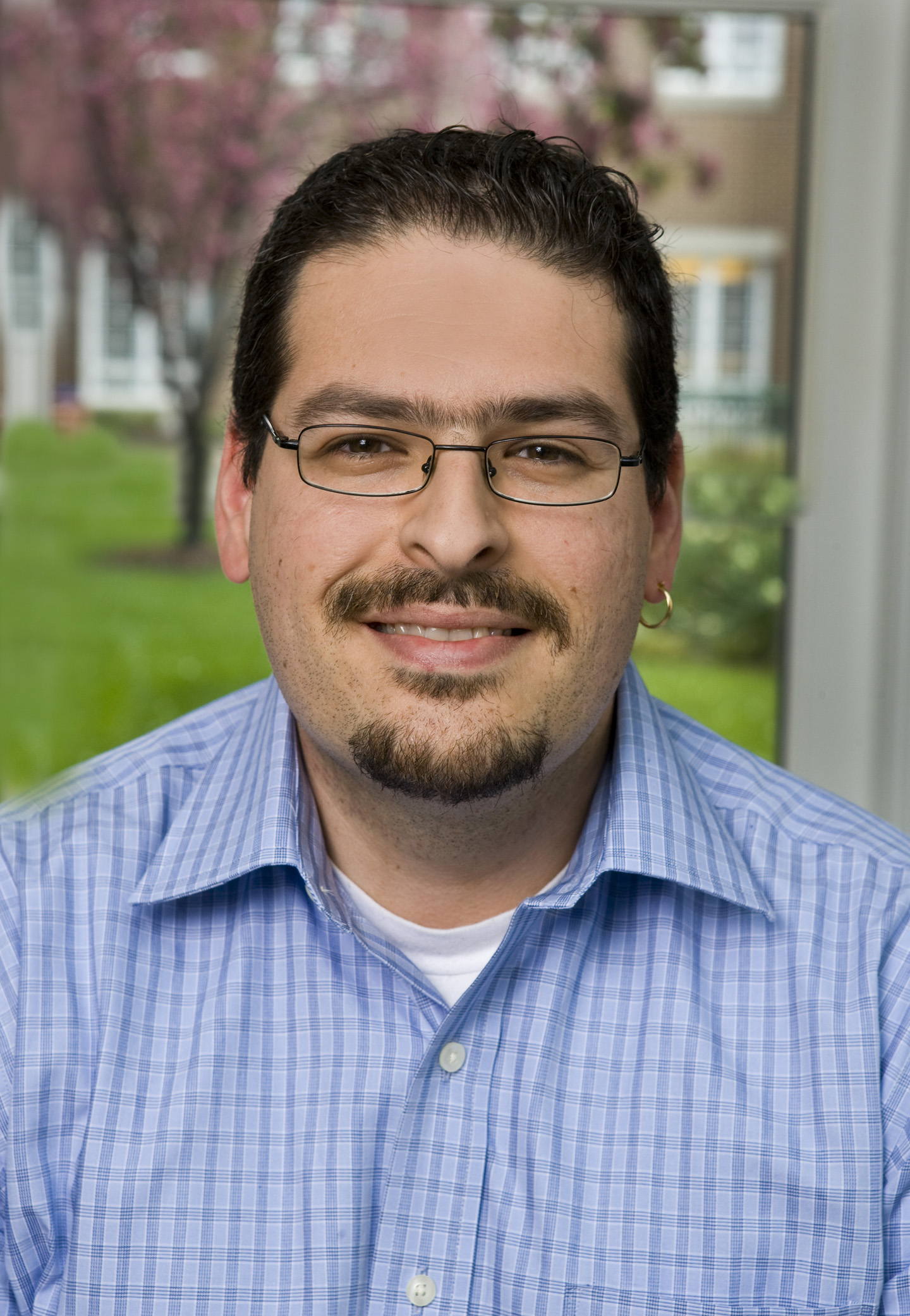
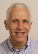
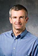
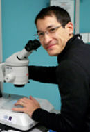
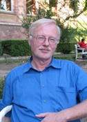
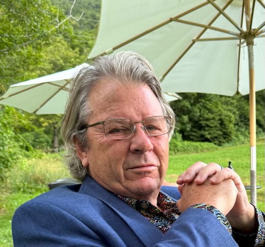
George Phillips
Rice University
Published May 31, 2025
For a person intent on tiny forms, George Phillips's career has been closely tied to big initiatives that have added new categories of structures to the Protein Data Bank. He has pioneered dynamic crystallography studies that reveal dramatic split-second moments of molecular activity. And his team has animated the atomic handbook on myoglobin, the well-studied molecular oxygen reservoir found in muscles.
But his most-cited paper remains a side note to the major themes of his laboratory. It happened almost by chance as a young faculty member at Rice University in Houston, Texas.
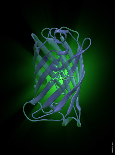
"That was actually kind of a one-off thing," Phillips says. "One of my college roommates called me and said, 'Hey, nobody's done the structure of green fluorescent protein. Do you want to work on it?' It crystallized easily, and we saw the structure of the native protein."
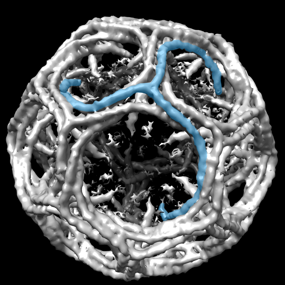
It was a cylinder shape, a beta sheet wrapped around an alpha helix. "We called it the beta-can," he says. "It was like Popeye's can of spinach." (Nature Biotechnology, 1996)
Fast-forward three decades, and Phillips may be one of the few US researchers in early 2025 whose future is not in jeopardy from the threat of massive cuts in federal funding that are looming over the US biomedical infrastructure and becoming a harsh reality for many. That's because Phillips is about to retire. Again.
Phillips earned his PhD at Rice and returned to start a new lab after a stint at the University of Illinois-Urbana Champaign. Then he spent a dozen years at University of Wisconsin-Madison, leaving with professor emeritus status when he returned to Rice in 2012.
Now Phillips is in the last few months of running his Rice lab, a decision made three years ago. He'll keep his office for a final foundation-funded project to tinker with a new way to solve the "phase problem" that bedevils X-ray crystallography.
Phase information is crucial to reconstruct the arrangements of atoms in a molecule, but it cannot be measured in the diffraction waves from a crystal, and so it is missing from every experimental data set. It's a problem that needs to be solved to visualize any molecule analyzed by X-ray crystallography.
"The classical methods take a lot of skilled crystallographer time, and we want to see if we can help that process along by training a neural network to shortcut the problem," he says.
The computational approach was inspired by the way AlphaFold cracked the protein folding problem. He, colleague Anastasios Kyrillidis and graduate student Tom Pan showed proof of concept on simple dipeptides and have since worked their way up from two amino acids to tackle molecules composed of 50–150 amino acids.
The project highlights a theme underlying Phillips's life in science. He seeks to make the technical chores more efficient, so that structural biologists and crystallographers can "spend more of their brain power to find interesting problems or think about what they mean," he says.
He views each of the two sub-specialties as the antidote for the other when things get boring. "A structural biologist, even if they're using the technique of crystallography, is primarily motivated by getting the structure and explaining what the structure means. Crystallographers are motivated by understanding the science of crystallography and advancing the art of crystallography for its own sake … and are mostly agnostic about what they're studying."
In his crystallography role, he has helped lead federally funded long-term projects that have advanced the tools and techniques for structural biology.
Unknown Proteins
In one, Phillips and his UWM colleagues won funding to establish one of the centers of the Protein Structure Initiative. The initiative was part of an international effort in structural proteomics.
"Structural biologists had looked around, and the structures that were being solved were things that had been studied in the lab already, and one wanted a reductionist explanation of how they worked," he says. "The [new] idea was, instead of going after the known proteins, let's go after the 'unknowneome,' we called it, tongue in cheek."
Combining expertise in X-ray crystallography (Phillips), nuclear magnetic resonance (John Markley) and Arabidopsis transcriptomics (Mike Sussman), the center took on the only eukaryotic organism in the initiative. Their charge was to go after structures of proteins with unknown functions and to automate the procedures for efficient high-throughput crystallography.
The larger initiative churned out more than 600 experimental structures every year for 10 years, assisted by new software, cloning procedures, and high throughput protein crystallization robots. Phillips's main responsibility at the UW center was "to oversee the crystallographic operation, everything from quality control on the protein through the crystallization, the screening of the crystals, the collecting of data at the synchrotron, the solving of the structure and the publishing of the work," he says.
Many times, initiative researchers found, even when an amino acid sequence looked like nothing anyone had seen, the protein structure wound up resembling a known protein. That led to the possibility of a kind of reverse structural biology, where researchers could test if the unknown doppelgänger had a similar biological function. In the last few years of the initiative, Phillips and other research partners put these new technologies to use in the discovery of novel enzymes found in natural product pathways to explore their pharmaceutical potential.
Laser! Detector! Action!
In another initiative just winding down now, Phillips teamed up with a consortium working on the next generation of structural biology - to capture molecules in motion, rather than the usual static snapshots. Launched in 2013, the BioXFEL Science and Technology Center featured the first X-ray free-electron laser (XFEL) on the planet based at Stanford University's linear accelerator facility. It was about a billion times brighter than other sources. Since then, a half dozen more such state-of-the-art lasers in facilities around the world have been set up, he says.
They took an approach called time-resolved crystallography to the next level. One of Phillips's jobs was to develop the right crystals for the system and to connect the results to meaningful biological questions. Other team members focused on how to design jets to shoot a focused stream of crystals and how to process the diffraction data.
"It takes a lot of protein to do these experiments," he notes. "An X-ray free electron laser produces rays that are so intense that your crystal is vaporized more or less instantly," Phillips says. "It's called serial femtosecond crystallography - serial, because you have to have a fresh crystal each time, and femtosecond, because that's the length of the pulse."
In BioXFEL, Phillips also helped pioneer another technique of catching split-second conformational changes within crystals. Many enzymes remain active in crystal samples and may have room to wiggle if no wild motions are required. Each batch of crystals can be mixed with a molecular partner for different periods of time before being streamed and zapped. "With different time delays, you take your crystallographic data, and then you can solve the structures of steps along the way, and then play them like a movie," he says.
Using this technique, for example, Phillips and his colleagues studied an enzyme produced by bacteria to defend themselves against antibiotics. Thanks to XFEL, they watched the enzyme bind to the antibiotic and saw the chemical changes as the enzyme degraded it. (Structural Dynamics, 2016; Nature Communications, 2023)
New ways of advancing scientific knowledge are great, but the consortium projects also satisfied the educator in Phillips. Early in his first Rice tenure, he wrote an online program for teaching crystallography basics, called X-RayView (https://xrayview.org/). It now also works on smartphones. For BioXFEL, he rebooted the educational component, serving as education and diversity director for eight years, which included hosting undergraduate students with diverse backgrounds in a summer research opportunity program.
He believes in giving back to the scientific community in other ways. He is editor-in-chief of the journal Structural Dynamics and past president of the American Crystallographic Association.
Early Days
Phillips was born in Dallas, the son of an airline pilot (father) and flight attendant (mother) who resigned as required when they married. His dad had been a transport pilot in World War II and the Korean War. The family relocated to Marin County, near San Francisco, where his dad worked for an airline chartered to fly US troops and supplies back and forth to Vietnam.
Science and math came easily to Phillips in high school in California. He returned to Texas for college at Rice in Houston. At a chemistry department open house for undergraduates, he wandered into a lab where students and postdocs were building atomic models. They were stacking plastic sheets to put together a three-dimensional representation of a chemical compound. This was before computer graphics, and it looked like they were playing with Tinker Toys.
They told him it was called crystallography, and it was kind of like solving puzzles to learn more about the chemical compound. "Wait. You can get paid to solve puzzles and play with Tinker Toys? I was all over that," says Phillips, whose office is decorated with modern colorful 3D printed plastic molecular models.
When he was a senior, Phillips came face-to-face with protein crystallography for the first time. A biochemistry professor, Florante Quiocho, had just grown some crystals of a bacterial binding protein and badly wanted a student to solve the structure. He convinced Phillips to leapfrog his pending undergraduate graduation and simultaneously begin his PhD project. He did a postdoctoral fellowship at University of Illinois, where he studied muscle regulatory proteins.
Animating myoglobin
Soon after he started his first lab at Rice, Phillips began what became a long collaboration with colleague John Olson, who was studying myoglobin, an oxygen-storing protein found in muscles, as a model for hemoglobin, the oxygen-transporting protein in blood.
Myoglobin was the first protein to have its three-dimensional structure solved in 1958. The static structure revealed the oxygen was trapped and it needed to move to function. Several generations of scientists since then used available techniques to explore potential conformational changes. Controversy ensued about how oxygen gets in and out.
"I did a lot of work with John on myoglobin trying to understand how the oxygen and other ligands get in and out of the protein," Phillips says. Long before BioXFEL, he came up with the idea of making a movie to see the action. His team used a orange laser beam to knock carbon monoxide (an experimental surrogate for oxygen) off the iron atom and watch where it went. "It rattled around and stuck again," he says. But that rattle also triggered some other movements that led the team to suggest a histidine gate may open as a part of its function. (Science, 2003)
A very stable version of myoglobin enables whales and other deep-diving mammals to produce high concentrations of the protein before it acquires the oxygen, the team discovered more recently. As a result, whales can pack 10–20 times more myoglobin into their muscles than humans and explains why their meat is so dark.
As he winds down his lab work, Phillips is turning his administrative and technical skills to a holistic garden plot at Rice. Students provide the labor, and they get to eat the fruits of their farming at university food services. He looks forward to tilling soil, pulling weeds, and maybe even fixing the tractor. In the meantime, he's using a lower-tech laser to etch paving bricks with donor names as a fundraiser for the garden. "It's very relaxing."
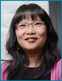
-Carol Cruzan MortonOther tales
-
![]()
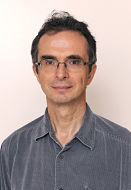
Drugging the Undruggable
Dhirendra Simanshu
Published 29 June 2026
![]()
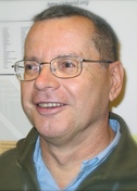
Structure-guided Antibiotics
Andrés Palencia
Published 27 March 2026
-
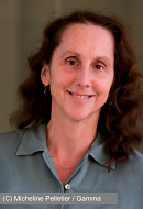
![]()
The Stories Antibodies Tell
Jean-Philippe Julien
Published 27 January 2026
![]()
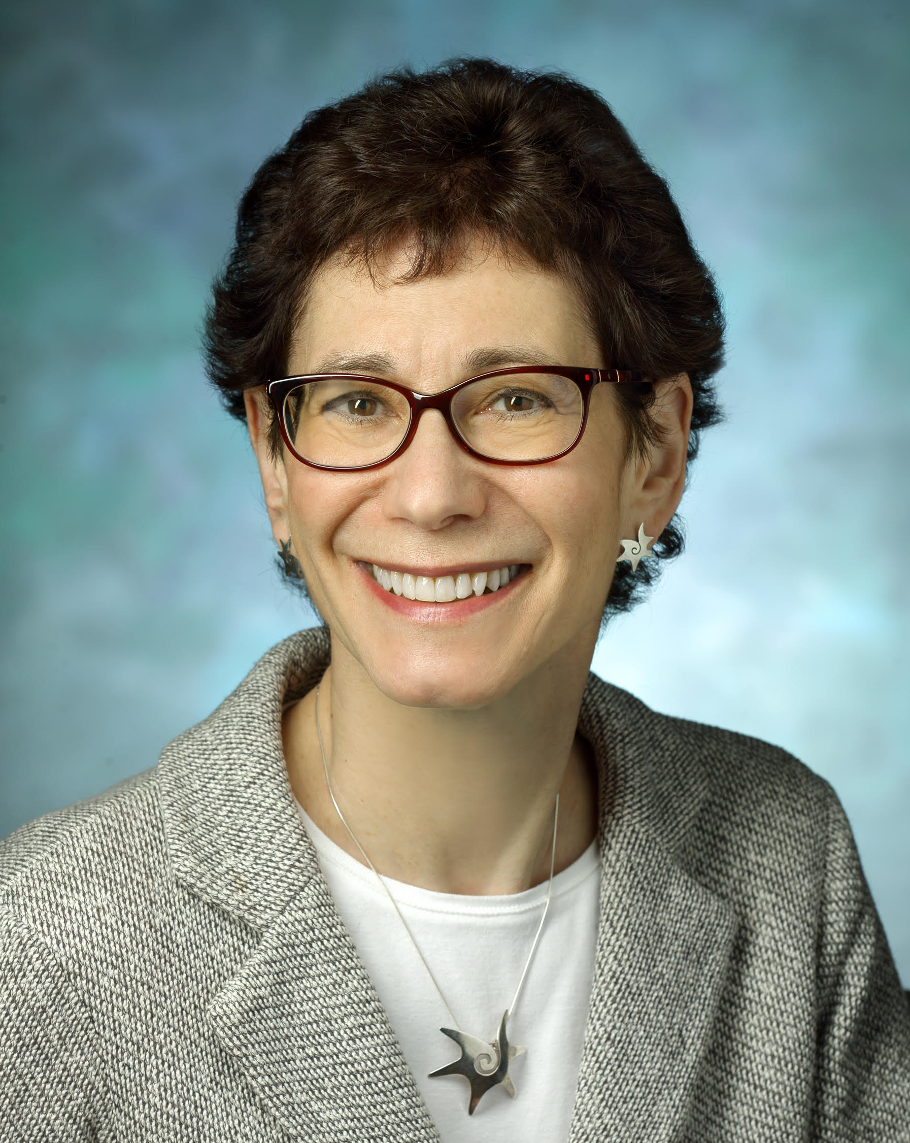
Reshaping Membranes
Melanie Ohi
Published 23 November 2025
-
![]()
Probing Microbes
Gira Bhabha
Published 30 September 2025
![]()
Drawn to the Light
Emina Stojković
Published 30 July 2025
-
![]()
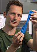
The Final Phase
George Phillips
Published 31 May 2025
![]()
Mind and Muscle
Ryan Hibbs
Published 28 March 2025
-
![]()
The Shapes of Energy
Luke Chao
Published 12 December 2024
![]()
Predicting Proteins
Jens Meiler
Published 25 November 2024
-
![]()
Death Metal
Steven Damo
Published 28 April 2024
![]()
Context Matters
Bing Chen
Published 30 January 2024
-
![]()
The Crystal Whisperer
Sarah Bowman
Published 29 November 2023
![]()
Data in Motion
Nozomi Ando
Published 29 September 2023
-
![]()
The Monstrous Maw
André Hoelz
Published 28 June 2023
![]()
Second Takes
Andrea Thorn
Published 28 February 2023
-
![]()
Radical reactions
Yvain Nicolet
Published 31 January 2023
![]()
Floppy Physics
Eva Nogales
Published 30 November 2022
-
![]()
Structure of Equity
Jamaine Davis
Published 28 September 2022
![]()
Life and Death of a Cell
Evris Gavathiotis
Published 28 July 2022
-
![]()
Follow the glow
Kurt Krause
Published 29 April 2022
![]()
Resolution solutions
Willy Wriggers
Published 25 February 2022
-
![]()
Of enzymes and membranes
Ming Zhou
Published 28 October 2021
![]()
Step-by-step
Gabrielle Rudenko
Published 26 September 2021
-
![]()
Moving muscle
Montserrat Samso
Published 26 July 2021
![]()
Particle catcher
Stefan Raunser
Published 28 June 2021
-
![]()
Designer drugs
Ho Leung Ng
Published 25 February 2021
![]()
Right place, right time
Ernesto Fuentes
Published 29 January 2021
-
![]()
Shape-shifting secrets of membranes
James Hurley
Published 27 November 2020
![]()
Enzymatic action
Cynthia Wolberger
Published 28 September 2020
-
![]()
Rules of motion
Priyamvada Acharya
Published 31 July 2020
![]()
Cosmic Squared
Michael Cianfrocco
Published 27 June 2020
-
![]()
Kaps are Cool
Yuh Min Chook
Published 28 April 2020
![]()
Spiraling into focus
Carsten Sachse
Published 29 March 2020
-
![]()
Seeing cilia
Alan Brown
Published 27 February 2020
![]()
For the Love of EM
Guy Schoehn
Published 27 January 2020
-
![]()
Protein Puddles
Michael Rosen
Published 16 December 2019
![]()
Changing channels
Daniel Minor Jr.
Published 27 September 2019
-
![]()
Listening Tips
Marcos Sotomayor
Published 30 July 2019
![]()
Beyond Cool
Published 31 May 2019
-
![]()
Hao Wu
A Higher Order
Published 30 May 2019
![]()
Aye Aye Captain
Alexandre Bonvin
Published 29 April 2019
-
![]()
The PARP Family Family
John Pascal
Published 28 February 2019
![]()
Frame by frame
Nikolaus Grigorieff
Published 28 January 2019
-
![]()
Predicting Success
Bil Clemons
Published 18 December 2018
![]()
Curiouser and Curiouser
Ramaswamy Subramanian
Published 27 November 2018
-
![]()
Rely on This
Sjors Scheres
Published 26 October 2018
![]()
Proteins out of bounds
Gerhard Wagner
Published 27 September 2018
-
![]()
Hiding in plain sight
Gaya Amarasinghe
Published 27 July 2018
![]()
Jumping Genes
Orsolya Barabas
Published 27 June 2018
-
![]()
Data Whisperer
Karolin Luger
Published 30 May 2018
![]()
Flipping the Switch
Jacqueline Cherfils
Published 27 April 2018
-
![]()
Tooling Around
Andrew Kruse
Published 29 March 2018
![]()
Comings and Goings
Tom Rapoport, Ph.D.
Published 23 February 2018
-
![]()
Transcriptional Rhythm
Seth Darst
Published 27 January 2018
![]()
The Language of Gene Regulation
Daniel Panne
Published 21 November 2017
-
![]()
Not Your Average Protein
James Fraser
Published 23 October 2017
![]()
Message Received
Sebastien Granier
Published 24 August 2017
-
![]()
Resistance is Futile
Celia Schiffer
Published 28 July 2017
![]()
Twist of Fate
Leemor Joshua-Tor
Published 28 June 2017
-
![]()
Drug Designer
John Buolamwini
Published 30 May 2017
![]()
Mathematically Minded
James Holton
Published 28 April 2017
-
![]()
Garbage Out
Kay Diederichs
Published 30 March 2017
![]()
Fixer Upper
Brandt Eichman
Published 27 February 2017
-
![]()
Mobilizers
Phoebe Rice
Published 31 January 2017
![]()
Escape Artist
Katya Heldwein
Published 19 December 2016
-
![]()
Nature’s Confectioner
Jochen Zimmer
Published 29 November 2016
![]()
State of Fusion
Jason McLellan
Published 27 October 2016
-
![]()
Here Be Dragons
Brian Fox
Published 28 September 2016
![]()
SBGrid Assumes Ownership of PyMOLWiki
Published 15 September 2016
-
![]()
Pharm Team
Oleg Tsodikov
Published 24 August 2016
![]()
Spiro-Gyra
Alejandro Buschiazzo
Published 27 July 2016
-
![]()
Turning the DIALS
Nicholas Sauter
Published 29 June 2016
![]()
Pipeline Dreams
Bridget Carragher and Clint Potter
Published 26 April 2016
-
![]()
U-Store-It
The SBGrid Data Bank provides an affordable and sustainable way to preserve and share structural biology data
Published 28 March 2016
![]()
Big Questions, Big Answers
Jennifer Doudna
Published 22 February 2016
-
![]()
Not a Structural Biologist
Enrico Di Cera
Published 17 December 2015
![]()
Divide and Conquer
Kevin Corbett
Published 19 November 2015
-
![]()
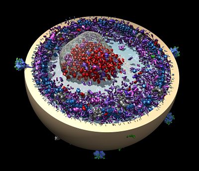
Computing Cellular Clockworks
Klaus Schulten
Published 23 October 2015
![]()
Trans-Plant
Gang Dong
Published 26 September 2015
-
![]()
Keep on Moving
James Berger
Published 23 August 2015
![]()
Totally Tubular
Antonina Roll-Mecak
Published 27 July 2015
-
![]()
From Disorder, Function
Julie Forman-Kay
Published 29 June 2015
![]()
Into Alignment
Geoff Barton
Published 27 May 2015
-
![]()
Two Labs, Many Methods
Michael Sattler
Published 28 April 2015
![]()
Picture This
Georgios Skiniotis
Published 20 March 2015
-
![]()
Intron Intrigue
Navtej Toor
Published 20 February 2015
![]()
Cut and Paste
Martin Jinek
Published 28 January 2015
-
![]()
Basics and Beyond
Qing Fan
Published 18 December 2014
![]()
Bloodletting and Other Studies
Pedro José Barbosa Pereira
Published 25 November 2014
-
![]()
Wire Models, Wired
A brief history of UCSF Chimera
Published 29 October 2014
![]()
In Search of…New Drugs
Doug Daniels
Published 30 September 2014
-
![]()
An Affinity for Affinity…and Corals
John C. Williams
Published 29 August 2014
![]()
Pete Meyer, Ph.D.
Research Computing Specialist
Published 22 August 2014
-
![]()
Justin O'Connor
Sr. System Administrator
Published 20 August 2014
![]()
Carol Herre
Software Release Engineer
Published 15 August 2014
-
![]()
Elizabeth Dougherty
Science Writer
Published 13 August 2014
![]()
Andrew Morin, Ph.D.
Policy Research Fellow
Published 11 August 2014
-
![]()
Jason Key, Ph.D.
Associate Director of Technology and Innovation
Published 8 August 2014
![]()
Piotr Sliz, Ph.D.
Principal Investigator, SBGrid
Published 1 August 2014
-
![]()
New Kid on the Block
James Chen
Published 29 July 2014
![]()
Membrane Master
Tamir Gonen
Published 30 June 2014
-
![]()
The Natural Bridge
Piotr Sliz
Published 13 June 2014
![]()
Surprise, Surprise
Catherine Drennan
Published 26 April 2014
-
![]()
Gone Viral
Olve Peersen
Published 20 March 2014
![]()
All Who Wander Are Not Lost
Frank Delaglio
Published 24 February 2014
-
![]()
The Raw and the Cooked
Graeme Winter
Published 24 January 2014
![]()
Vacc-elerator
Peter Kwong
Published 17 December 2013
-
![]()
Structural Storyteller
Karin Reinisch
Published 15 November 2013
![]()
The Fixer
Jane Richardson
Published 28 October 2013
-
![]()
Inside the Box
Mishtu Dey
Published 17 September 2013
![]()
Sensing a Change
Brian Crane
Published 16 August 2013
-
![]()
Towards Personalized Oncology
Mark Lemmon
Published 16 July 2013
![]()
Brush with Fame
Yizhi Jane Tao
Published 14 June 2013
-
![]()
Toxic Avenger
Borden Lacy
Published 21 May 2013
![]()
Pushing the Boundaries
Stephen Harrison
Published 22 April 2013
-
![]()
Strength in Numbers
Joseph Ho
Published 18 March 2013
![]()
One Lab, Many Methods
Wesley Sundquist
Published 12 February 2013
-
![]()
Unplanned Pioneer
Tim Stevens
Published 15 January 2013
![]()
From Actin to Action
Emil Pai
Published 11 January 2013
-
![]()
Unstructured
A Brief History of CCP4
Published 12 December 2012
![]()
Stop, Collaborate and Listen
Eleanor Dodson
Published 5 November 2012
-
![]()
X-PLORer
Axel Brunger
Published 1 October 2012
![]()
Share the Wealth
Zbyszek Otwinowski
Published 22 August 2012
-
![]()
Unraveling RNA
Anna Pyle
Published 18 July 2012
![]()
Sharper Image
Pawel Penczek and SPARX
Published 4 June 2012
-
![]()
Creative Copy Cat
Pamela Bjorkman
Published 25 April 2012
![]()
Charm and Diplomacy
Gerard Kleywegt
Published 7 March 2012
-
![]()
From Curiosity to Cure
Marc Kvansakul
Published 13 December 2011
![]()
The Lure of the Sandbox
Paul Emsley and Coot
Published 15 October 2011
-
![]()
Springsteen, Tolkien, Protein
Alwyn Jones and Frodo
Published 17 June 2011
![]()
Structures Solved Simply
Paul Adams and Tom Terwilliger on Phenix
Published 2 June 2011
-
![]()
Escape from the Darkroom
Wolfgang Kabsch and XDS
Published 19 May 2011
![]()
Playing the Odds
Randy Read and Phaser
Published 19 May 2011
-
![]()
Crystallography for Kids
Lynne Howell
Published 17 May 2011
![]()
Better, Faster, Stronger, More
Victor Lamzin and ARP/wARP
Published 17 May 2011